Examination of the genitalia should occur within the context of a complete physical exam. Children generally respond well to this part of the examination when the examiner is confident, uses age-appropriate language to explain what is happening, and can find ways to engage the child in conversation.
Using a Colposcope
In some medical centers, a colposcope is available and can be utilized in the examination. The advantages of a colposcope include an excellent light source, magnified view of the external anatomy, and opportunity to record the findings of the examinations when the colposcope is attached to a camera. It can be particularly helpful in children because of its nonthreatening and noninvasive characteristics.
Alternatives to a Colposcope
An otoscope with the earpiece removed is an adequate substitute for magnification and can be used as an alternative. Some centers are using digital cameras with macro lenses for close up viewing and creating images of the external genitalia. If this is appropriately explained to the patient, and the photographer is willing to testify regarding the image, this may also be appropriate. For more information on photography, see DOCUMENTATION: Photographic Documentation.
Positioning
The supine position is most commonly used in genital exams, particularly in the pre-adolescent population. The supine lithotomy position with stirrups is reserved for cooperative female adolescents. Younger children are placed in the supine frog leg position on the parent's lap or exam table. Placing a child in various positions will assist in the exam.
The prone knee chest position is most commonly used for the perineal and anal examinations because it frequently affords an optimal view of the posterior hymen and/or the vagina, thus revealing the cervix and trauma or a foreign body. When using the prone knee chest position, the medical provider and assistant should guide the child into the position and provide constant reassurance. When examining a child who has been abused in the knee chest position, be sensitive to the potential emotional discomfort when the child assumes this position for exam purposes. Placing the child in the lateral knee chest position (lateral decubitus) or standing leaning over the examination table for an anal examination may be less emotionally traumatic.
Inspection/Palpation of the Genitalia
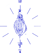
Document findings of the anal/genital region on anatomical drawings, noting location and the explanation offered for any lesions found. Always document the exam position when indicating the lesions on the anatomic drawings. A superimposed clock face can assist in accurate documentation for females (Figure A). Conventionally, "12 o'clock" is anterior.
Figure A: Clock face superimposed on female genital area
Reprinted from the New York State Department of Health Child and Adolescent Sexual Offense Medical Protocol.
If within 96 hours of the sexual assault, examine for dried or moist secretions, ecchymotic grab marks, bite marks, or for evidence of other injuries. Note any healed scars, STD lesions, or other abnormal findings.
Perineum
Note the presence of any fresh or healed lacerations, STD lesions, pubic lice, rashes, or other unusual findings.
Vaginal or urethral discharge
Note presence of discharge in terms of amount, color, and presence of odor. Identify source, the vagina or urethra.
Female Genital Examination
The genital examination of the prepubertal child is principally external visualization. Instrumentation is rarely necessary. All visualization should be done before attempting any specimen collection. Never use an adult size speculum to examine a prepubescent child. The examination of the pubertal child may require the use of a vaginal speculum, particularly if the adolescent was previously sexually active. If the use of a speculum is required, use the smallest size possible.
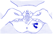
Labial traction is a technique to assist in the examination of the female genitalia. This technique can be used to open the vaginal orifice to better inspect the vagina for trauma, foreign body, and discharge and the hymenal anatomy for tears, scars, and attenuation. To use the labial traction technique in the supine frog leg position, grasp the labia gently between the thumb and index finger of each hand, and exert gentle downward and lateral traction (Figure B). Do not insert fingers to determine the size of the hymenal opening or its patency.
Figure B: Labial traction in the supine frog leg position
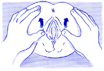
Figure C: Labial traction in the prone knee chest position
Reprinted from the New York State Department of Health Child and Adolescent Sexual Offense Medical Protocol.
When the child is in the prone knee chest position, separating the labia with both hands will display the hymen and vagina (Figure C). This position may give better visualization of the posterior rim of the hymen as it is pulled downward by gravity. If this causes such discomfort that the child is unable to cooperate, use another position. One alternative is to have the child lie supine, holding her knees to her chest.
If visualization of the hymen remains inadequate, it can be enhanced by using a moistened Q-tip to shift redundant tissue if the child is pubertal or by irrigating the hymen with saline if the child is prepubertal. Medical providers with expertise in the Foley catheter balloon technique have also used this examination technique.
Gently retract the labia majora to observe the genital structures. If the child is relaxed, the vagina and introitus will gradually open to reveal its maximum size. The size of the vaginal opening is affected by position and the degree of the relaxation/sedation of the child. For this reason, the exam position and degree of relaxation should be documented in the medical record when noting the transverse vaginal opening size. Variation exists in the hymenal anatomy configuration among nonabused children.
Examine the following:
Labia majora and minora
Note any skin lesions, unusual pigmentations, or other skin changes.
Clitoris
Note unusual size or changes of the clitoris or hood.
Urethral meatus
Note any signs of inflammation, edema, or other lesions of the periurethral tissue.
Perihymenal tissue (vestibule)
Note any increases in vascularity, abrasions, lacerations, scarring, or STD lesions.
Hymen
Note the configuration (annular, redundant/fimbriated, crescentic, dorsal/anterior) of the hymen on the anatomic drawing. Observe for signs of trauma or STD lesions. Evaluate for significant distortion of the hymenal shape, fresh tears, transsections, fresh hemorrhages, abrasions, and bruises. In addition, evaluate for ecchymotic areas, healed scars or adhesion, rounded or thickened edges, attenuated edges or loss of hymenal volume, and abnormal vascular pattern. Note decreases in hymenal tissue and assess whether any absence of tissue represents a shallow notch, a deep notch, or a complete transsection. The terms "intact, broken, virginal, marital, or missing" are not sufficient to describe hymenal findings.
Posterior fourchette and fossa navicularis
Note lacerations or scars, bruises, healing abraded areas, STD lesions, or neovascularization.
Vagina
Note any bleeding, discharge, STD lesions, foreign bodies, abnormal vascular pattern, petechiae, or other lesions on the walls of the vagina.
Cervix
Note any bleeding, discharge, STD lesions, cervicitis, tears, or other signs of trauma.
Male Genital Examination
Examine the following:
Penis
Note whether patient is circumcised. Note any STD lesions, bite marks, edema, hematomas, lacerations, abrasions, or dried secretions.
Urethral Meatus
Note any scars, STD lesions, discharge, or bleeding.
Scrotum
Note any erythema, ecchymoses, STD lesions, abrasions, or bite marks.
Testes
Note the presence of descended testes, any signs of atrophy or differential in firmness of the tissue.
Anal/Rectal Examination
With either the supine or prone knee-chest position, the examiner should use both hands to separate the buttocks for viewing of the anal area in males or females. In general, traction techniques are not necessary when these positions are used. Note in the medical record the exam position that is utilized and the degree of relaxation.
The anus is readily visualized in the prone knee chest position. Because this position may cause some victims unusual embarrassment or recall memories of prior abuse, two other positions that may be more acceptable are the lateral decubitus (knee chest) position and standing bending forward over the exam table. In the lateral decubitus position, the child should be curled so that the knees are as close to the chest as possible, usually with one leg flexed more than the other. The examiner can separate the buttocks with one hand on each gluteal area, using the thumb and fingers for leverage.
Examine the following:
Buttocks
Note fresh or healed lesions, dried secretions, ecchymoses, rashes, STD lesions, handprints or fingerprints.
Perianal skin
Examine for presence of inflammation. Record findings of dried secretions, bruising, tears, lacerations, fissures, tears, or lacerations that are located on the external surface, internal to the sphincter, or extend across the pectinate line, which is the juncture between the anal mucosa and the anal epithelium.
Anal verge/folds/rugae
Note whether the verge or anal sphincter skin folds appear to be prominent, normal, or flattened (funneled) when the child is in a relaxed state.
Tone
Note whether anal tone is within normal limits. A visual examination is almost always sufficient to assess anal tone. Avoid a digital rectal examination in children unless there is suspicion of internal trauma. On visual exam, note the presence of anal spasm.
Anal laxity
Estimate or measure the diameter of any anal dilatation. Note the length of time to reach maximum dilatation and whether the dilatation was constant or intermittent. Apply only enough lateral traction to separate the buttocks. An anus that dilates to greater than 15 millimeters in diameter with gentle buttocks traction and without stool present is a significant observation. Record the presence or absence of stool in the rectal ampulla.
Rectal examination
A rectal examination is rarely necessary in the evaluation of sexual abuse. In situations where significant intraabdominal or rectal trauma is suspected, or when the presence of a foreign body is suspected, a digital examination is justified. Consider further evaluation utilizing anoscopy and possible sedation. In situations following an acute assault involving trauma to the rectum, perform a guaiac exam.